
RTG 3
Imaging
Radim Chmelík (CEITEC), Nicolas Blanc (ETH), Michal Stöger-Pollach (TUW)
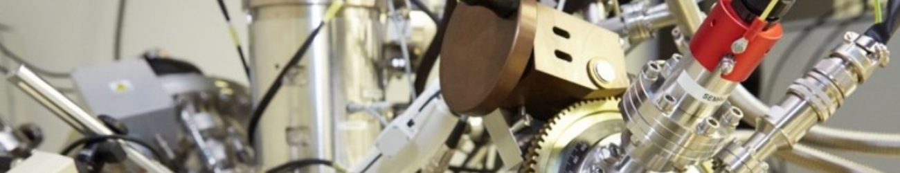
Observing catalytic reaction by in-situ real-time microscopy
Not so long ago, a single chemical reaction saved humanity from food shortages. It was the catalytic reaction of ammonia synthesis using the Haber-Bosch process. After the discovery of this reaction, the world population grows according to the amount of ammonia produced. Today, humanity is facing a shortage of planet-friendly energy sources rather than a shortage of food. Catalytic reactions could help to solve the problem again. Despite the fact that today we have advanced analytical methods, catalytic reactions are still not well understood, even the most studied ones such as the oxidation of carbon monoxide to carbon dioxide. This reaction has applications in car catalysts but is also important in the removal of CO from the hydrogen production process, which is one of the alternatives for closed renewable energy cycles. It is important that existing analytical methods are improved and ideally measure the progress of the reaction in real-time and at different scales. Therefore, we started to observe catalytic reactions using electron and ion microscopes in real-time, and our aim is to make our observations of these reactions useful for a more thorough understanding of them and subsequently for their mass application in the sustainable energy industry, similarly, as Haber and Bosch did in the first half of the last century in the case of the catalytic synthesis of ammonia.
STEM-EELS in plasmonics
This group focuses on a branch of nanophotonics called plasmonics which studies localized surface plasmon resonances (LSPR, where plasmon is the effect of interaction of electromagnetic field with electrons). A characteristic feature of LSPR is a strong enhancement of the electromagnetic field in the surrounding material together with its spatial confinement on the subwavelength scale in the vicinity of plasmonic antennas, which can be utilized in a wide range of applications including spectroscopy and sensing, energy harvesting, and medicine. Significance of LSPR is further increased by easy tunability of their spectral characteristics via engineering the size, shape, or dielectric environment of nanoparticles. Mapping of LSPR modes with high spatial and energy resolution is necessary to understand their nature and properties and to be able to design plasmonic antennas with a better performance in applications. Scanning transmission electron microscopy (STEM) combined with electron energy loss spectroscopy (EELS) has become a favorite technique to map LSPR with a nanometer-scale spatial and better than 0.1 eV energy resolution. We have developed a technique based on Babinet’s principle to study the magnetic near field in the plasmonic antenna, which enables us to map also plasmonic antennas with magnetic hot spots and design them more efficiently for the local magnetic field enhancement. Thus we are able to develop absorption spectroscopy of magnetic transitions for the study of rare-earth ions in the visible region or electron paramagnetic resonance with higher sensitivity.
Optical microscopy
An optical microscopy is an unmatched tool in science and real-life practical applications ranging from bio-imaging to technical fields. The current progress of microscopy drives the development of new light shaping methods overcoming the performance of standard optical components made of glass. Using liquid crystal or metal nanostructures enables the fabrication of physically thin optical components shaping the light through the transformation of its polarization state instead of the optical path. We build on the potential of these new-generation components, which are beneficial thanks to polarization-dependent properties, lightweight design, and their simple integration into the existing optical systems. It enables the development of new optical modalities, which we successfully deploy to study artificial nanostructures (plasmonic nanoantennas), colloidal nanoparticles made of noble metals, or even naturally created biological photonic structures. Developed imaging techniques aim to become helpful in the fabrication processes of plasmonic nanostructures, biosensing, or the study of the live cells and their interactions with colloidal nanoparticles.